


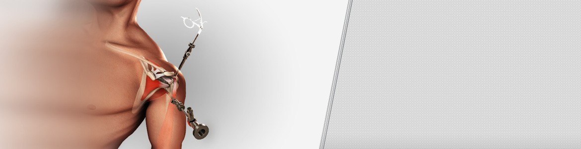


Bankart Tear
The shoulder joint (glenohumeral joint) is a ball and socket joint, where the head of the upper arm bone (humerus) attaches to the shoulder socket (glenoid cavity). The shoulder socket is extremely shallow and therefore needs additional support to keep the shoulder bones from dislocating. The labrum, a cuff of cartilage that encircles the shoulder socket, helps serve this purpose by forming a cup for the humeral head to move within. It provides stability to the joint, enabling a wide range of movements.
The labrum can sometimes tear during a shoulder injury. A specific type of labral tear that occurs when the shoulder dislocates is called a Bankart tear. This is a tear to a part of the labrum called the inferior glenohumeral ligament and is common in younger patients who sustain a dislocation of the shoulder. A Bankart tear makes the shoulder prone to repeat dislocation in patients under 30 years of age.
Diagnosis
Your physician will ask about your medical history and perform a thorough physical examination of your shoulder. Your doctor may recommend additional studies such as X-ray’s or an MRI.
Treatment
Conservative treatment measures for a bankart tear include rest and immobilization with a sling followed by physical therapy.
Bankart repair surgery is indicated for a bankart tear when conservative treatment measures do not improve the condition, but instead results in repeated shoulder joint dislocation.
Description of Procedure
Bankart surgery can be performed using an open surgical technique or a minimally invasive surgical technique known as arthroscopy.
Open Surgery
You will be placed in a chair inclined at a 30-degree angle with your arm positioned freely over the end of the operating table.
Your surgeon makes a long incision over the shoulder joint and cuts through the soft tissues in order to gain access to the interior of the joint. Retractors are used to hold the tissue apart so the surgeon can view the detachment of the labrum from the glenoid socket. Small holes are drilled through the edge of the glenoid socket and anchors are inserted into the area of the tear. These anchors have suture threads attached to them. These suture threads are then passed through the holes to reattach the torn labrum securely to the joint capsule. At the end of the procedure the incision is closed and dressings are applied.
Arthroscopic Procedure
During an arthroscopic Bankart procedure, your surgeon makes a few small incisions over your shoulder joint. An arthroscope, a slender tubular device attached with a light and a small video camera at the end is inserted through one of the incisions into your shoulder joint. The video camera transmits the image of the inside of your shoulder joint onto a television monitor for your surgeon to view. Your surgeon then uses small surgical instruments through the other tiny incisions to trim the edges of your glenoid cavity. Suture anchors are then inserted to reattach the detached labrum to the glenoid. The tiny incisions are then closed and covered with a bandage.
Arthroscopy causes minimal disruption to the other shoulder structures and does not require your surgeon to detach and reattach the overlying shoulder muscle (subscapularis) as with the open technique.
Post-operative Care
- After your surgery, you will spend about an hour in the recovery room.
- Your physical therapist will start you on shoulder exercises the day following your surgery to strengthen and improve the range of motion of your shoulder joint.
- You will be allowed to perform your daily activities as tolerated, but without lifting objects heavier than a plate or glass while you heal.
- Your arm may be placed in a sling for three weeks to restrict use of your operated shoulder.
- You may resume light low-risk activities, like jogging and swimming 8 to 10 weeks after surgery, and may be advised to avoid contact sports for some time.
Risks
BANKART repair is a relatively safe procedure but as with any surgical procedure potential risks and complications may occur. Some of the potential risks associated include:
- Infection
- Injury to adjacent nerves or blood vessels
- Stiffness of the joint
- Pain
- Persistent instability
- Need for additional surgeries